Imagine spending two decades with a debilitating growth in your mouth. With a protruding tumour on one jaw. Your face is disfigured. Drugs are only enough to keep you alive and pain is the only thing you feel.
This has been the case for Pendo Masonga, a Tanzanian national who has battled a tumour on her left jaw for years on end. But now, the mother of one can breathe easy, thanks to oral and maxillofacial surgery (OMFS) that she underwent recently.
Last week, a team of medics led by Cuban Oral and Maxillofacial Surgeon, Dr Arelis Rabelo Castillo, conducted an eight-hour surgery at the Kitui County Referral Hospital to remove a 500g malignant growth that has been giving Pendo sleepless nights.
Oral and maxillofacial surgery is no mean feat. Ideally, a patient would check into a hospital, see a doctor and when the diagnosis necessitates an operation, get an appointment with a surgeon at the theatre.
This is not usually the case with OMFS procedures. More often than not, they are done as emergency operations.
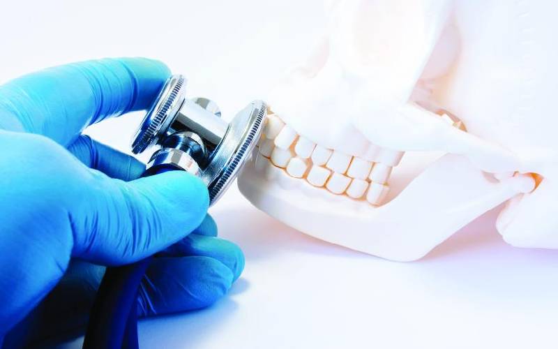
Think of the oral and maxillofacial region of your body like the mouth and all its connecting regions. This is the area around your face, head and neck. An oral and maxillofacial surgery, as the name suggests, is a surgical speciality that deals with reconstructive surgery of this region.
Data from the Kenya National Bureau of Statistics show that 4,672 people were seriously injured in road accidents in 2018. Out of 2,827 people who were killed in road accidents that year, the National Transport and Safety Authority (NTSA) reported that 511 were motorcyclists. It is such accidents that are the leading causes of maxillofacial trauma.
Sports injuries, falls, physical assaults and burns are other common causes of maxillofacial injury. Patients classified as having maxillofacial trauma can present with a variety of injuries, ranging from minor to life-threatening. This kind of injury affects one’s ability to breath, speak or eat.
Oral and maxillofacial surgeons have to be trained both as dentists then as surgeons to enable them to first correctly diagnose oral diseases after which they will be equipped with surgical skills. Typically, they do more emergencies than other surgeons and are highly trained to treat a wide spectrum of diseases, injuries and defects.
OMFS is used to treat problems such as facial deformity and misaligned jaws, tumours and cysts of the jaw, mouth ulcers, head and neck cancer and skin cancer. Despite the increasing number of injuries caused by road accidents, Kenya has just about 31 such specialists against a population of over 47 million.
How it is done
Doctors start by doing imaging tests to determine if cancer has spread beyond the mouth. The stage of cancer will determine which treatment option will be pursued.
Small cancers can be removed through minor surgery but in special cases like Pendo’s, the procedure involves removing a section of your jawbone after which a reconstructive surgery will be done to rebuild your mouth to help you regain the ability to talk and eat.
Because this procedure is done under general anaesthesia, the patient needs at least two days to recover. The OMFS is majorly a reconstructive procedure and shouldn’t be confused with other cosmetic procedures.
Stay informed. Subscribe to our newsletter
3D printing in oral and maxillofacial surgery
3D printing is being used in multiple applications in medicine and industry and is now being adapted for use in oral and maxillofacial surgery. 3D printing has been used to design titanium plates and other materials to accurately maintain or rebuild accurate reproductions of the lost bony structures. This helps save on time and costs.
Digital impressions
To reproduce the structures of the oral cavity, it requires a dental impression captured using material placed inside the mouth. A stone material is then poured in the impression to produce a stone copy of the oral tissues. Breakthroughs in technology have made it possible to digitally capture images of the structures of the oral cavity, and transfer this into a digital format that can be used for multiple applications. A small wand placed in the mouth photographs the structures of the oral cavity and converts this into the digital form used to create the design needed for the prosthesis.
 The Standard Group Plc is a
multi-media organization with investments in media platforms spanning newspaper
print operations, television, radio broadcasting, digital and online services. The
Standard Group is recognized as a leading multi-media house in Kenya with a key
influence in matters of national and international interest.


